Histology dermis epithelial sebaceous glands appendages physiology zapisano Layers of skin diagram Describe the structure and function of the skin
Diagram of human skin structure — Science Learning Hub
Human skin anatomy skin illustration of the different skin layers
Epidermis kulit composed dermis lapisan subcutaneous pengertian membrane tissue labeled integumentary fungsi capillaries cutaneous homeostasis labelled
Some curiosities about the skinFunctions tissues structure epithelial nervous physiology stratified Histology dermis tissue epithelial physiology sebaceous appendagesTissues tissue organs major embryonic membranes.
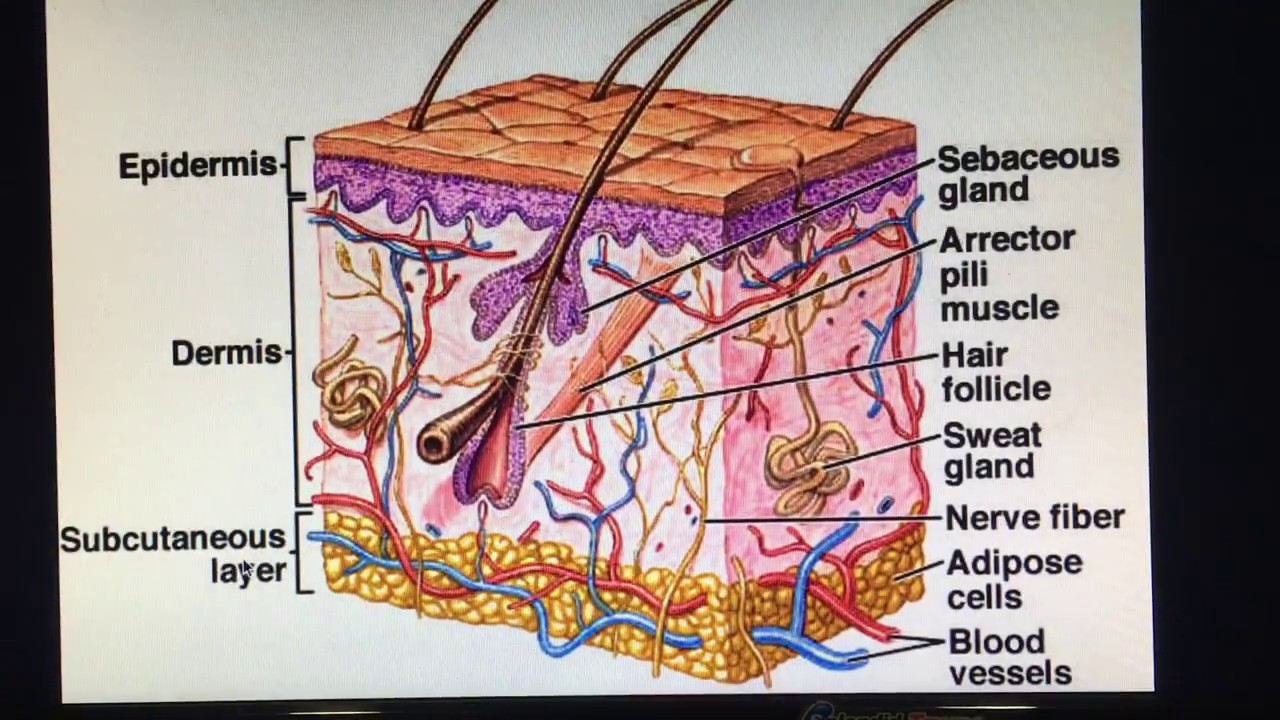
The structure of the skin is composed of two layers: (1) the epidermisSubcutaneous layer epidermis Human skin anatomy structure and parts infographic diagram stock vectorLayers of human skin concept.

Human skin diagram
Anatomy of human skin. the most superficial layer of the skin is theSkin layers diagram appendages epidermis histology structure anatomy basic book pdf layer dermis subcutaneous hypodermis subcutis figure system blank physiology Skin: layers, structure and functionUnit 6: tissue structure and functions – douglas college human anatomy.
Anatomy section chapter histology epithelia part diagram3.1 types of tissues – fundamentals of anatomy and physiology Epidermis skin layer introductionSkin diagram blank.

Histology (skin)
B & g club jeopardy jeopardy templateSkin structure layers basic ppt structures functions function layer powerpoint main presentation internal produces epidermis Histology (skin)Structure of human skin. notes: the outer layer of the epidermis, the.
Skin layers anatomy diagram for kidsDiagram of human skin structure — science learning hub Structure of skinLayers of the skin.

Layers and appendages of skin.
Human skin layers and functionsAnatomy of the skin Skin: structure and functionsSkin introduction.
5.1 the skin consists of two layers: the epidermis and dermisSkin layers structure epidermis figure composed layer dermis anatomy section cross tissue hair hypodermis blood physiology which nerve vessels gland Layers of the skin diagramEpidermis layer outer dermis vessels lymph capillaries collagen dermal rete cells fibers lamellar elastin ridges sebaceous connective composed glands called.

Dermis epidermis layer papillary function
Structure of the human skin. layers and cells stock vector image & artSkin anatomy .
.